Erasmus MC heeft ervoor gezorgd dat je Mijn BSL eenvoudig en snel kunt raadplegen. Je kunt je links eenvoudig registreren. Met deze gegevens kun je thuis, of waar ook ter wereld toegang krijgen tot Mijn BSL.
Om ook buiten de locaties van Erasmus MC, thuis bijvoorbeeld, van Mijn BSL gebruik te kunnen maken, moet je jezelf eenmalig registreren. Dit kan alleen vanaf een computer op een van de locaties van Erasmus MC.
Eenmaal geregistreerd kun je thuis of waar ook ter wereld onbeperkt toegang krijgen tot Mijn BSL.
Login
Als u al geregistreerd bent, hoeft u alleen maar in te loggen om onbeperkt toegang te krijgen tot Mijn BSL.
The online version of this article (doi:10.1007/s12471-015-0756-8) contains supplementary material, which is available to authorized users.
B.C. Du Pré, L.W. Van Laake contributed equally to this manuscript.
A 32-year-old woman presented with progressive dyspnoea for 6 weeks, fatigue, weight loss, chest pain, and night sweats. Apart from an uncomplicated delivery of her third child 4 months ago, she had no medical or family history. Pneumonia was suspected (chest X-ray Supplementary Fig.1a, b), but thoracic computed tomography (CT) was performed to exclude pulmonary emboli.
The CT surprisingly showed a large intrathoracic mass of 94 × 80 mm extending from the sternum to the thoracic spine, compressing the superior and inferior caval veins, the right atrium, and (partly) the right ventricle (Fig. 1, Supplementary Fig. c–e, and Video 1). Further analysis revealed a giant coronary aneurysm (GCA) originating from the proximal right coronary artery (RCA) connecting inferiorly to the distal RCA. Left ventricular function was good (Video 2 and 3). Due to the size and the location of the GCA, surgical exclusion was the treatment of choice (Fig. 2, Supplementary Fig. F). After surgery, the patient recovered well and was discharged on the fifth postoperative day.
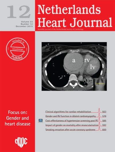
Fig. 1
Thoracic CT showing a large intrathoracic mass. Note the equal densities of the aneurysm and the left ventricular lumen
Fig. 2
Giant coronary aneurysm after sternotomy
×
×
GCAs are extremely rare: less than 0.02 % of all cardiac surgery is attributed to GCAs [1]. They are usually related to comorbidities or injuries such as infectious disease, inflammatory disease, trauma, coronary angioplasty, or connective tissue disease but can also occur as a congenital abnormality. [4] It is likely that in the current case, the GCA had existed for many years. Increased workload and hormonal changes during pregnancy and delivery may have contributed to growth and symptoms of the GCA [2, 3].
Conflict of interest
None declared
Open Access This article is distributed under the terms of the Creative Commons Attribution License which permits any use, distribution, and reproduction in any medium, provided the original author(s) and the source are credited.
Het Netherlands Heart Journal wordt uitgegeven in samenwerking met de Nederlandse Vereniging voor Cardiologie. Het tijdschrift is Engelstalig en wordt gratis beschikbaa ...
